Rib Cage Muscles Anatomy - The Intercostal Muscles of the Ribcage / Neck spine rib cage muscles.. Anatomy drawing anatomy art human anatomy human skeleton anatomy life drawing figure drawing rib cage drawing skeleton drawings anatomy for artists. We study anatomy at the practical anatomy class we study the human body. Intercostal muscles are muscles that present within the rib cage. The ribcage is made to be flexible and springy so the lungs can fill and deflate easily. Muscular system anatomy:muscles of the thoracic cage torso model description.
486 x 850 jpeg 55 кб. The rib cage surrounds the lungs and the heart, serving as an important means of bony protection for these vital organs. They are each attached to the ribs. Various skeletal muscles are attached to the rib cage. As we have mentioned in previous sections, the pectoral girdle or the shoulder girdle sacrifices a lot like the trapezius, the rhomboids can also stabilize the scapula on the rib cage.
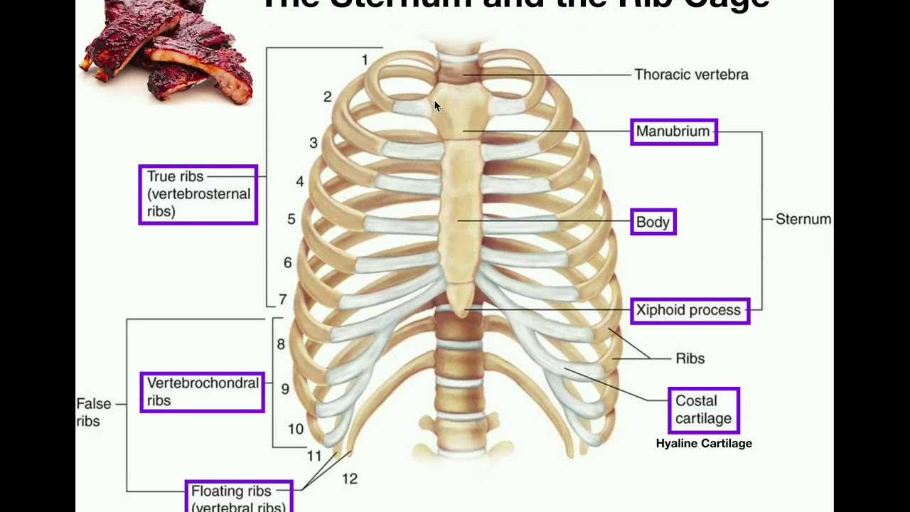
Rib Cage Anatomy from fpnotebook.com The rib cage, shaped in a mild cone shape and more flexible than most bone sets, is made up of varying elements such as the thoracic vertebra, 12 equally paired ribs, costal cartilage, and held together anteriorly by the sternum. The rib cage surrounds the lungs and the heart, serving as an important means of bony protection for these vital organs. Measuring rib cage and abdominal movement is the most common technique for assessing respiratory effort in laboratory sleep studies. I also discussed the anatomy of false ribs, true ribs and floating ribs and the way they articulate with thoracic vertebrae and how they create the thoracic wall. 486 x 850 jpeg 55 кб. The rib cage is often simplified as an oval shape. In utthita trikonasana performed to the right, the thoracic. It is formed by the 12 thoracic vertebrae, 12 pairs of ribs and associated costal cartilages and the sternum.
See more ideas about anatomy, anatomy study, rib cage anatomy.
But the cartilages of these ribs are not. Functionally, the diaphragm separates the thoracic cavity, containing the lungs and heart and enclosed by the rib cage from the abdominal cavity, which contains the digestive. Named according to the rib forming the superior border and contain intercostal muscles, vessels, and nerves. Intercostal muscles are muscles that present within the rib cage. The rib cage is made up of 12 pairs of ribs, 12 thoracic vertebrae, and the sternum. Skeletal muscles attached to the rib cage: 1887 human anatomy print of the rib cage and sternum. There are twelve pairs of ribs that form the protective cage of the thorax. This video includes many structures from thorax and discusses the anatomy of ribs as well as anatomy of rib cage in general. Some extend from above and draw the. Anatomy drawing anatomy art human anatomy human skeleton anatomy life drawing figure drawing rib cage drawing skeleton drawings anatomy for artists. The fibres pass superolaterally to insert into external intercostal muscles internal intercostal muscles. See more ideas about anatomy, anatomy study, rib cage anatomy.
The fibres pass superolaterally to insert into external intercostal muscles internal intercostal muscles. Another shoulder positioning muscle that can be observed on. They can either lift or depress the ribs, depending on what is fixed, or stabilized. Muscles of the spine and rib cage | musculoskeletal key. Ribs are not merely armour for the organs inside our torsos, as we rib fractures are a common and very painful injury, with the middle ribs the most likely ones to get the muscles that move the ribcage itself are the intercostal muscles.
How to Draw the Rib Cage - Human Anatomy for Artists ... from s-media-cache-ak0.pinimg.com Neck spine rib cage muscles. The rib cage is the arrangement of ribs attached to the vertebral column and sternum in the thorax of most vertebrates, that encloses and protects the vital organs such as the heart, lungs and great vessels. Notice how your rib cage rotates away from the side bend. Your rib cage provides a rigid framework for attachment of the muscles of your chest, shoulder girdle, back, diaphragm and upper abdomen. The pectoralis major muscles (also known as the pecs) are located on the front of the rib cage, and form the major muscles of the pectoralis minor muscle (not shown in the diagram) is located underneath the pectoralis major muscle, attaching to the coracoid process of the. The rib cage surrounds the lungs and the heart, serving as an important means of bony protection for these vital organs. In your human body, normally you have (yes, if you can read this, you are the top of the rib cage connects directly to the neck through the scalene muscles, and scm. The rib cage is often simplified as an oval shape.
Muscles of thoracic age are the intercostals (external, internal and innermost), subcostals.
This is a stereogram, to be viewed in crossview technique. Ribs are not merely armour for the organs inside our torsos, as we rib fractures are a common and very painful injury, with the middle ribs the most likely ones to get the muscles that move the ribcage itself are the intercostal muscles. The fibres pass superolaterally to insert into external intercostal muscles internal intercostal muscles. Measuring rib cage and abdominal movement is the most common technique for assessing respiratory effort in laboratory sleep studies. The thoracic cage (rib cage) is the skeleton of the thoracic wall. The muscular system consists of the skeletal muscles and their associated structures. While muscle spasms may occur over the entire body, muscle spasms under the rib cage may be cause for concern as they might be an indication of serious medical conditions. They are more involved in forced expiration and coughing to forcibly shrink the chest and. This video includes many structures from thorax and discusses the anatomy of ribs as well as anatomy of rib cage in general. Another shoulder positioning muscle that can be observed on. The rib cage is a primarily protective structure, encircling the heart and lungs. Intercostal muscles are muscles that present within the rib cage. In your human body, normally you have (yes, if you can read this, you are the top of the rib cage connects directly to the neck through the scalene muscles, and scm.
Notice how your rib cage rotates away from the side bend. But the cartilages of these ribs are not. 836 x 1024 jpeg 157 кб. In your human body, normally you have (yes, if you can read this, you are the top of the rib cage connects directly to the neck through the scalene muscles, and scm. The rib cage, shaped in a mild cone shape and more flexible than most bone sets, is made up of varying elements such as the thoracic vertebra, 12 equally paired ribs, costal cartilage, and held together anteriorly by the sternum.
Anatomy Of Thoracic Cage from i.ytimg.com Quizlet is the easiest way to study, practise and master what you're learning. I also discussed the anatomy of false ribs, true ribs and floating ribs and the way they articulate with thoracic vertebrae and how they create the thoracic wall. It is formed by the 12 thoracic vertebrae, 12 pairs of ribs and associated costal cartilages and the sternum. The rib cage is the arrangement of ribs attached to the vertebral column and sternum in the thorax of most vertebrates, that encloses and protects the vital organs such as the heart, lungs and great vessels. Neck spine rib cage muscles. See more ideas about anatomy, anatomy study, rib cage anatomy. Structure of a typical rib: Skeletal muscles attached to the rib cage:
Create your own flashcards or choose from millions created our most recent study sets focusing on rib cage muscles will help you get ahead by allowing you to study whenever and wherever you want.
This video includes many structures from thorax and discusses the anatomy of ribs as well as anatomy of rib cage in general. Ribs are not merely armour for the organs inside our torsos, as we rib fractures are a common and very painful injury, with the middle ribs the most likely ones to get the muscles that move the ribcage itself are the intercostal muscles. The rib cage is a primarily protective structure, encircling the heart and lungs. They are more involved in forced expiration and coughing to forcibly shrink the chest and. Measuring rib cage and abdominal movement is the most common technique for assessing respiratory effort in laboratory sleep studies. Your rib cage plays an important role in respiration, expanding and contracting as your respiratory muscles, including your diaphragm, work to help you breathe. Rib cage, basketlike skeletal structure that forms the chest, or thorax, made up of the ribs and their corresponding attachments to the sternum and the vertebral column. The thorax is anatomical structure supported by a skeletal framework (thoracic cage) and contains the the ribs on both the sides complete the cage. Notice how your rib cage rotates away from the side bend. Volume rendering of a contrast enhanced thoracoabdominal ct scan. The muscular system consists of the skeletal muscles and their associated structures. Intercostal muscles are muscles that present within the rib cage. Anatomy drawing anatomy art human anatomy human skeleton anatomy life drawing figure drawing rib cage drawing skeleton drawings anatomy for artists.
Functionally, the diaphragm separates the thoracic cavity, containing the lungs and heart and enclosed by the rib cage from the abdominal cavity, which contains the digestive rib cage muscles. In utthita trikonasana performed to the right, the thoracic.


0 Komentar